Single Molecule Imaging in Live Cells
Single Molecule Imaging in Live Cells


-
 1. Single molecule imaging in liv…
0
00:00/00:00
1. Single molecule imaging in liv…
0
00:00/00:00 -
 2. Untitled: Slide 2
144.41107774441107
00:00/00:00
2. Untitled: Slide 2
144.41107774441107
00:00/00:00 -
 3. Outline
287.88788788788787
00:00/00:00
3. Outline
287.88788788788787
00:00/00:00 -
 4. Three cell types of interest
354.22088755422089
00:00/00:00
4. Three cell types of interest
354.22088755422089
00:00/00:00 -
 5. Tissue cell
380.28028028028029
00:00/00:00
5. Tissue cell
380.28028028028029
00:00/00:00 -
 6. The Fluid Mosaic Model of the …
427.52752752752752
00:00/00:00
6. The Fluid Mosaic Model of the …
427.52752752752752
00:00/00:00 -
 7. Three different membrane archi…
530.93093093093091
00:00/00:00
7. Three different membrane archi…
530.93093093093091
00:00/00:00 -
 8. The E. coli outer envelope
648.78211544878218
00:00/00:00
8. The E. coli outer envelope
648.78211544878218
00:00/00:00 -
 9. Untitled: Slide 9
714.61461461461465
00:00/00:00
9. Untitled: Slide 9
714.61461461461465
00:00/00:00 -
 10. The labels we use
761.09442776109449
00:00/00:00
10. The labels we use
761.09442776109449
00:00/00:00 -
 11. Objective-type total internal …
889.82315648982319
00:00/00:00
11. Objective-type total internal …
889.82315648982319
00:00/00:00 -
 12. Imaging single molecules
957.05705705705714
00:00/00:00
12. Imaging single molecules
957.05705705705714
00:00/00:00 -
 13. Particle position determinatio…
1102.3690357023691
00:00/00:00
13. Particle position determinatio…
1102.3690357023691
00:00/00:00 -
 14. Outline
1131.5315315315315
00:00/00:00
14. Outline
1131.5315315315315
00:00/00:00 -
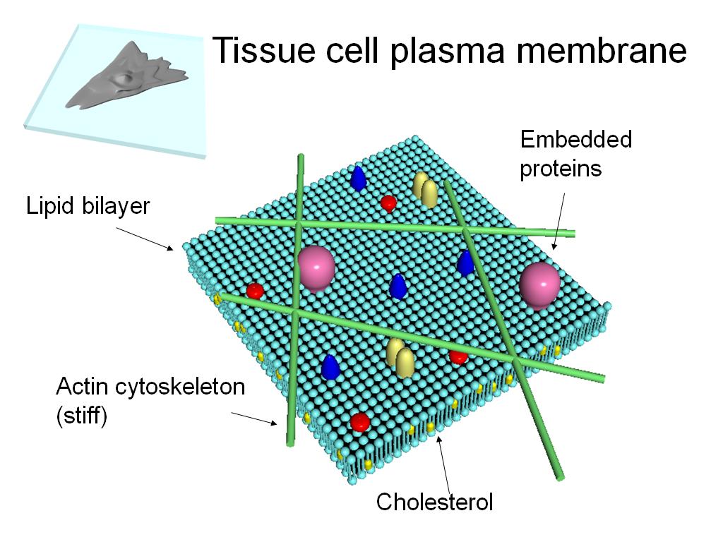 15. Tissue cell plasma membrane
1137.9379379379379
00:00/00:00
15. Tissue cell plasma membrane
1137.9379379379379
00:00/00:00 -
 16. Transferrin receptor on NRK ce…
1147.7811144477812
00:00/00:00
16. Transferrin receptor on NRK ce…
1147.7811144477812
00:00/00:00 -
 17. Takahiro Fujiwara, Akihiro Kus…
1218.6186186186187
00:00/00:00
17. Takahiro Fujiwara, Akihiro Kus…
1218.6186186186187
00:00/00:00 -
 18. Analysis of Diffusion
1302.6359693026361
00:00/00:00
18. Analysis of Diffusion
1302.6359693026361
00:00/00:00 -
 19. Membrane Skeleton Fence
1430.3636970303637
00:00/00:00
19. Membrane Skeleton Fence
1430.3636970303637
00:00/00:00 -
 20. Anchored-Protein "Picket" Mode…
1519.91991991992
00:00/00:00
20. Anchored-Protein "Picket" Mode…
1519.91991991992
00:00/00:00 -
 21. translational diffusion
1623.9906573239907
00:00/00:00
21. translational diffusion
1623.9906573239907
00:00/00:00 -
 22. Oligomerization-Induced Trappi…
1714.8148148148148
00:00/00:00
22. Oligomerization-Induced Trappi…
1714.8148148148148
00:00/00:00 -
 23. Outline
1799.7997997997998
00:00/00:00
23. Outline
1799.7997997997998
00:00/00:00 -
 24. RBC plasma membrane
1811.1444778111445
00:00/00:00
24. RBC plasma membrane
1811.1444778111445
00:00/00:00 -
 25. Spectrin and band 3
1867.3673673673675
00:00/00:00
25. Spectrin and band 3
1867.3673673673675
00:00/00:00 -
 26. DIDS Biotin linker
2035.6690023356691
00:00/00:00
26. DIDS Biotin linker
2035.6690023356691
00:00/00:00 -
 27. Video of a quantum dot
2079.8465131798466
00:00/00:00
27. Video of a quantum dot
2079.8465131798466
00:00/00:00 -
 28. Typical trajectories of quantu…
2107.1404738071406
00:00/00:00
28. Typical trajectories of quantu…
2107.1404738071406
00:00/00:00 -
 29. Red cell pathologies
2175.4754754754754
00:00/00:00
29. Red cell pathologies
2175.4754754754754
00:00/00:00 -
 30. Affect of hereditary anemias o…
2345.9125792459126
00:00/00:00
30. Affect of hereditary anemias o…
2345.9125792459126
00:00/00:00 -
 31. GC Kodippili et al. (2009) Blo…
2441.9419419419419
00:00/00:00
31. GC Kodippili et al. (2009) Blo…
2441.9419419419419
00:00/00:00 -
 32. Dmicro
2504.7380714047381
00:00/00:00
32. Dmicro
2504.7380714047381
00:00/00:00 -
 33. Outline
2648.7153820487156
00:00/00:00
33. Outline
2648.7153820487156
00:00/00:00 -
 34. Why E. coli?
2663.9973306639972
00:00/00:00
34. Why E. coli?
2663.9973306639972
00:00/00:00 -
 35. Outline
2679.6463129796466
00:00/00:00
35. Outline
2679.6463129796466
00:00/00:00 -
 36. Diffusion of GFP in the E. col…
2699.6329662996332
00:00/00:00
36. Diffusion of GFP in the E. col…
2699.6329662996332
00:00/00:00 -
 37. Diffusion of GFP in the E. col…
2785.9526192859526
00:00/00:00
37. Diffusion of GFP in the E. col…
2785.9526192859526
00:00/00:00 -
 38. Diffusion of GFP in the E. col…
2817.7510844177514
00:00/00:00
38. Diffusion of GFP in the E. col…
2817.7510844177514
00:00/00:00 -
 39. Diffusion of GFP in the E. col…
2857.8244911578245
00:00/00:00
39. Diffusion of GFP in the E. col…
2857.8244911578245
00:00/00:00 -
 40. Diffusion of GFP in the E. col…
2910.8775442108777
00:00/00:00
40. Diffusion of GFP in the E. col…
2910.8775442108777
00:00/00:00 -
 41. Outline
3041.3413413413414
00:00/00:00
41. Outline
3041.3413413413414
00:00/00:00