[Illinois] Advanced Materials Characterization Workshop 2012: AM-FM and Loss Tangent Imaging, Two Tools for Quant. Nanomechanical Property Mapping
Licensed under Creative Commons.
Category
Published on
Abstract
Amplitude-modulated Atomic Force Microscopy (AM-AFM), also known as tapping mode, is a proven, reliable and gentle imaging method with wide spread applications. Previously, the contrast in AM-AFM has been difficult to quantify. In this work, we introduce two new techniques that allow unambiguous interpretation of material properties.
 AM-FM imaging combines the features and benefits of normal tapping mode with quantitative and high sensitivity of frequency modulated (FM) mode. Briefly, the topographic feedback operates in AM mode while the second resonant mode drive frequency is adjusted to keep the phase at 90 degrees, on resonance. The FM image returns a quantitative value of the frequency shift that in turn depends on the sample stiffness and can be applied to a variety of physical models. Loss tangent imaging is a recently introduced quantitative technique that recasts the interpretation of phase imaging into one term that includes both the dissipated and stored energy of the tip sample interaction.
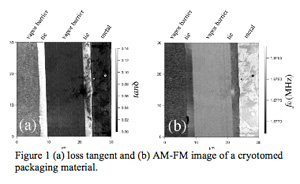
 These two quantitative techniques can be performed simultaneously. In Figure 1, a cryotomed, cross-sectioned area of a coffee bag packaging material has been imaged. The loss tangent image on the left clearly shows the highly lossy "tie" layers connecting the low-loss metal layer with two vapor-barrier polymer layers. The AM-FM image on the right shows the relative stiffness of the five layers, with the metal layer being the stiffest and the tie layers the softest. Figure 2 shows graphene deposited on a SiO2 substrate. The frequency channel shows clear contrast between the SiO2 and graphene layers. The loss tangent image shows that the boundary region between the SiO2 and graphene is dissipative.
Bio
Irene Revenko is an application scientist for Asylum Research since 2002. Prior to Asylum she was director of the Life Sciences Department at Veeco. Dr. Revenko received two Ph.D.s from Claude Bernard University, Lyon, France, one in 1998 on Applications of Atomic Force Microscopy in Biology and the other in 1994 on the Visualization of Type I Collagen Fibers.
Sponsored by
Platinum Sponsors: AIST-NT, Horiba Scientific, Micro Materials, Zeiss Asylum, Hysitron, Princeton Instruments
Sponsors: AFM Workshop, American Vacuum Society, Bruker, Chemplex, JEOL USA, Kurt J. Lesker Company, Olympus, Oxford Instruments, Thermo Scientific, Zygo, Agilent Technologies, Angstrom Scientific, Cameca, FEI Company, Kratos Analytical, Oerlikon Leybold Vacuum, Ophir Photonics, Panalytical, Witec
Cite this work
Researchers should cite this work as follows:
Time
Location
Engineering Sciences Building, University of Illinois at Urbana-Champaign, Urbana, IL
Submitter
NanoBio Node, Charlie Newman, Obaid Sarvana, AbderRahman N Sobh
University of Illinois at Urbana-Champaign